Macular telangiectasia type 2 (MacTel) is a disease of the retina, the light-sensing tissue at the back of the eye. MacTel leads to a gradual deterioration of central vision, which becomes noticeable to people around 50-60 years of age. The average age of diagnosis for MacTel patients is 57 years old.
The gradual loss of central vision people experience because of MacTel has a significant impact on quality of life. This is because central vision is required for tasks that require sharp vision, like reading and driving. In MacTel, the quality of a person’s central vision decreases over a period of 10 to 20 years, or more.
Macular telangiectasia type 2 is a bilateral disease, which means that both eyes are affected. However, the disease does not necessarily affect both eyes equally. It may progress more quickly in one eye than the other. A person with MacTel may see more clearly with one eye than the other. The disease may become apparent earlier in one eye than the other.

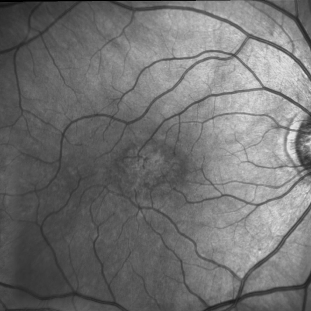
MacTel affects an oval-shaped area of the retina, centered on the fovea.
MacTel may be an inherited disease. The MacTel Project studies families with MacTel to determine the pattern of inheritance, and to look for responsible genes. Between 10-50% of an affected person’s first-degree relatives (parent, sibling, or child) may have MacTel. A “MacTel gene” has not yet been identified. In fact, multiple genes may be involved in the development of MacTel.
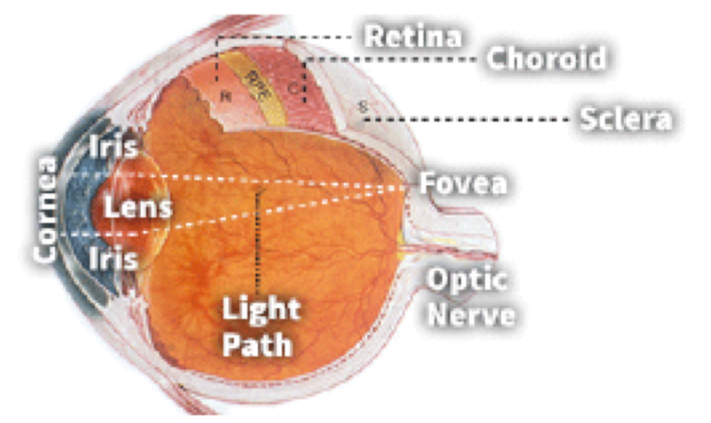
Macular telangiectasia type 2 affects a well-defined area of the retina, which is referred to as the “MacTel zone.” This is an oval-shaped area centered on the fovea. The fovea is contained within the macula, the central area of the retina. The fovea is the focal point for light entering the eye. It is the region of the retina with the highest density of cone photoreceptor cells. Humans rely heavily on the fovea to construct a clear picture of their surroundings. In fact, about half of the nerve fibers traveling from the retina to the brain carry information from the fovea. In the course of MacTel, photoreceptors in and around the fovea die.
The Lowy Medical Research Institute sponsored a Natural History Observation Study as part of the MacTel Project. This was the first study to examine health-related quality of life in MacTel patients. Results of the study showed that MacTel patients have reduced visual function compared to people without MacTel. Even though MacTel participants had measurable trouble with vision, standard visual acuity tests (i.e. reading the eye chart) did not always accurately reflect this.
The Lowy Medical Research Institute supports an Eye Donor program. Eyes donated from MacTel patients are used study the disease, leading to a better understanding of how MacTel affects the retina. In the MacTel eyes that have been donated for research, Müller glia cells were found to be missing from the MacTel zone. Müller glia cells play an important supportive role to photoreceptors in the retina. The cause of their disappearance from the MacTel zone in patients is still under investigation. Changes in the retinal pigment epithelial cells (RPE), and their function, are also apparent in the course of the disease. Clinically, there is a readily observed change in the retina’s blood vessels; they become dilated and take on new patterns. These physiological changes, and others, are the subject of LMRI research.


