Imaging

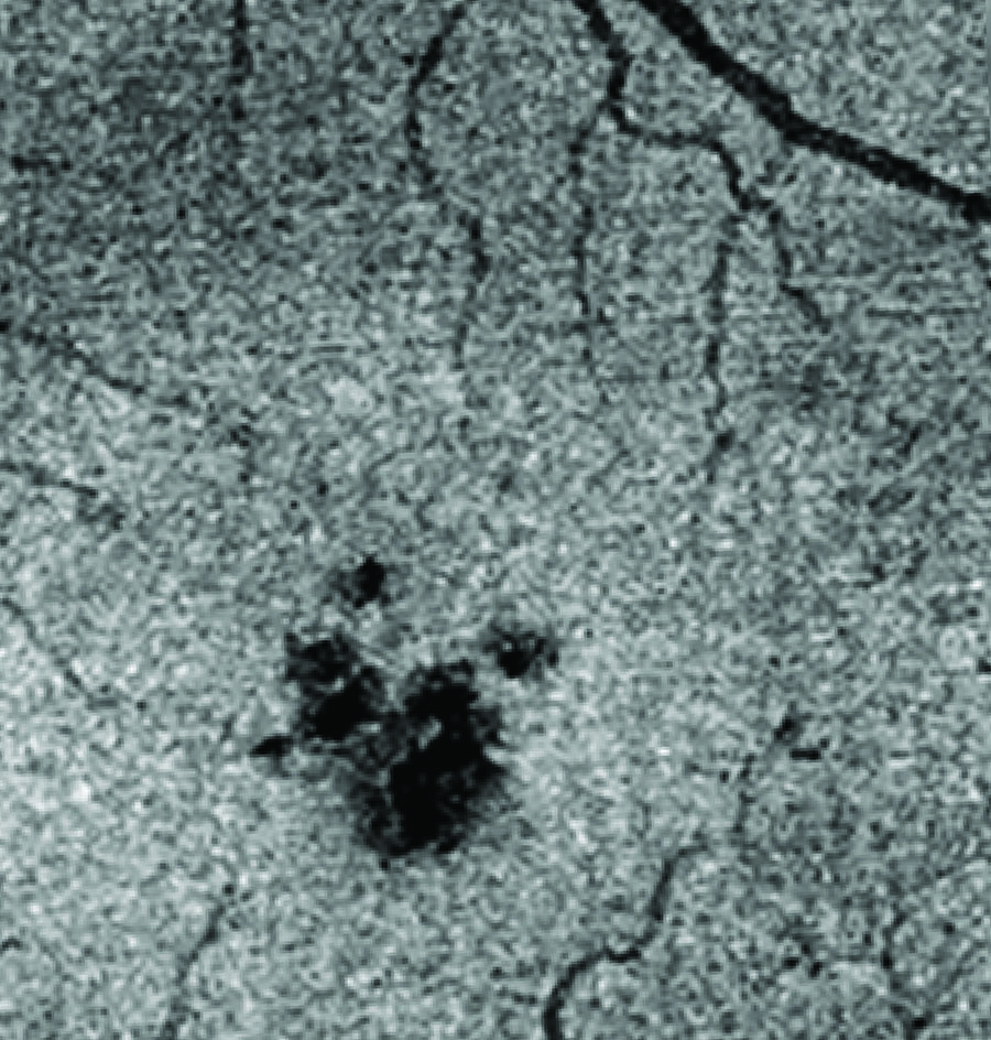
This en face OCT image of a MacTel eye shows a disruption in photoreceptors (dark patch) in the foveal region of the retina.

Advanced imaging technologies have changed our understanding of disease progression in macular telangiectasia type 2. Clinicians affiliated with the Lowy Medical Research Institute are using new imaging modalities to diagnose and follow changes in the MacTel eye.  Some of these imaging modalities were developed for research purposes, or are in early stages of development with manufacturers.  Many such specialized devices are not yet widely available across all LMRI-affiliated clinical sites.

Some of these technologies capture very high-resolution pictures of the retina, in some cases down to the cellular level.  For example, scientists can see individual photoreceptors by adaptive optics imaging.  Some clinicians who participate in the MacTel Project have specialized OCT devices that allow them to see blood vessels in the eye, with optical coherence tomography-angiography (OCT-A) machines.  Recent research indicates that the MacTel retina has a signature pattern observed by fluorescence lifetime imaging ophthalmoscopy (FLIO), which may allow for earlier diagnosis in the future, or diagnosis among family members.  While not widespread, such specialized imaging devices are very important to MacTel research.

Other imaging technologies, including optical coherence tomography (OCT) and microperimetry, are in use by members of the MacTel clinical community around the world.


Moorfields Reading Center

The Moorfields Reading Centre evaluates all ophthalmic images collected as part of the MacTel Project Registry and the Natural History Observation Study, and offers the official diagnosis of MacTel for patients who participate in these research projects.

The Moorfields Reading Centre for ophthalmic images was established by the Moorfields Eye Hospital NHS Foundation Trust, in partnership with the University College London Institute of Ophthalmology.  The Moorfields Reading Centre was established because there was a recognized need for a center that could create robust ophthalmic surveys and clinical trial project designs.  Clinical trials were hampered by an inability to develop robust outcome measures, in part because ophthalmic diseases had not been thoroughly characterized.  The Reading Centre serves as an unbiased, independent center for image analysis.

The Reading Centre standardizes the definition of diseased states, relating to the retina.  They also standardize what is classified as an abnormality in ophthalmic images.  They collect measurements from images, and grade images, so that changes can be recognized.

The classification of disease status has been extremely important to the MacTel Project.  One aspect of the MacTel Project is to analyze families with macular telangiectasia type 2.  Following the identification of one individual in a family with MacTel (the “proband”), their relatives may be examined for MacTel.  The Moorfields Reading Centre determines if family members are affected or unaffected.   This can be difficult for an age-related disease, where it is possible that an individual is not yet showing all of the symptoms of disease that would lead to a definitive diagnosis.

Duke Reading Center

The Duke Reading Center, located in North Carolina, is primarily a contract research center comprised of a group of ocular imaging, clinical trial, and biostatistics specialists.  The Duke Reading Center advises sponsors of ophthalmic clinical trials on the design and execution of their studies, reads ophthalmic images for clinical studies, and performs statistical analyses of image data.  They also certify and train study-site personnel in the capture and submission of ophthalmic images to the Reading Center according to study protocols.

The Duke Reading Center is the designated reading center for the NT-501 clinical trial.